05 / 05 · CT + scanDICOM folder + upper and/or lower scan
CT + scan
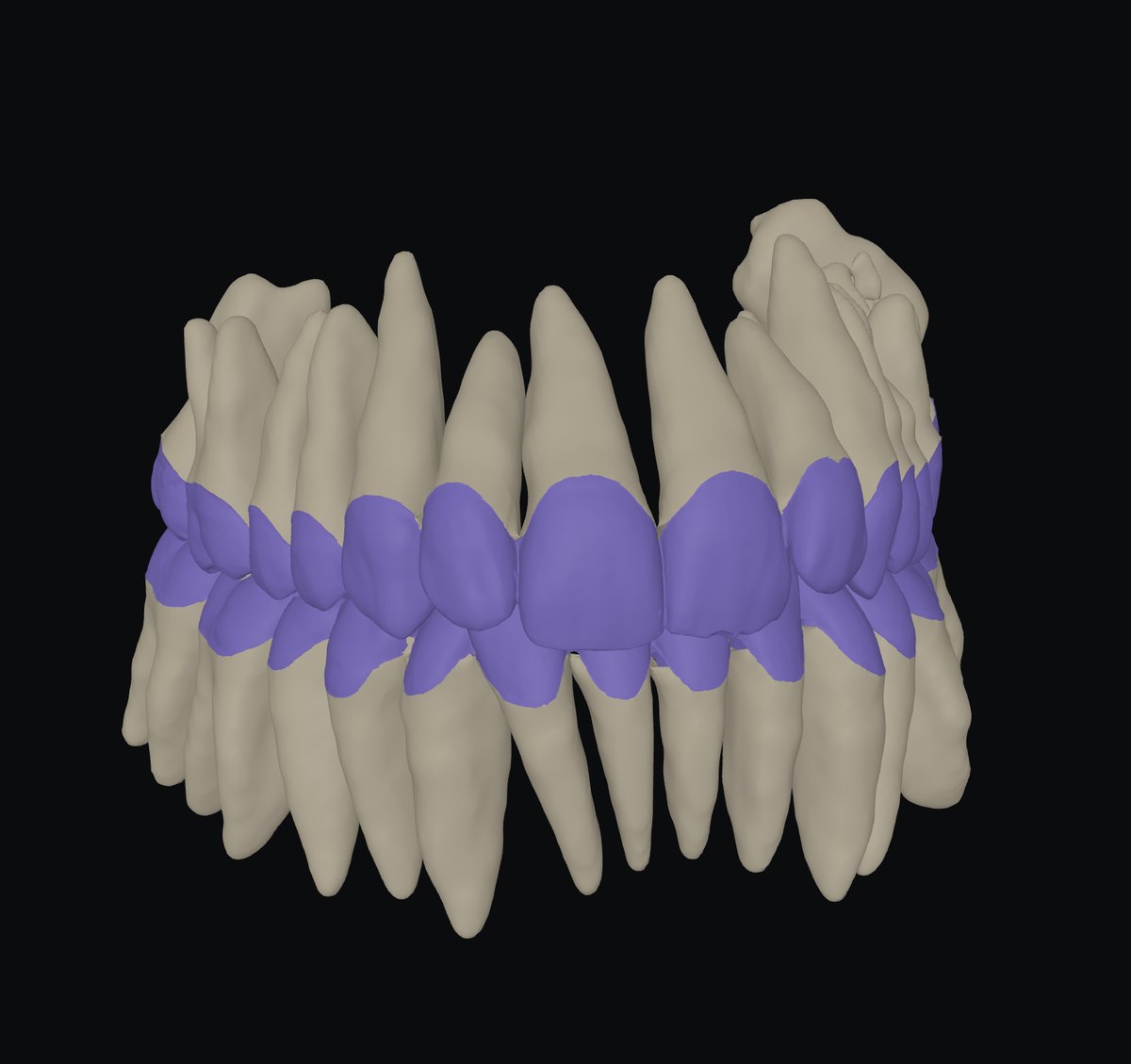
The jaw scans placed on the CT: every tooth a crown from the scan and a root from the CT.
An exact crown on a real root — for moving teeth, planning an implant or designing a bridge with the bone in view. No manual points.
InDICOM folder + scans
Out3D · STL · pose · link
RunsBrowser + server in the EU
KeptNothing
How to use it3 steps · on a real case
- 1CT + scans in
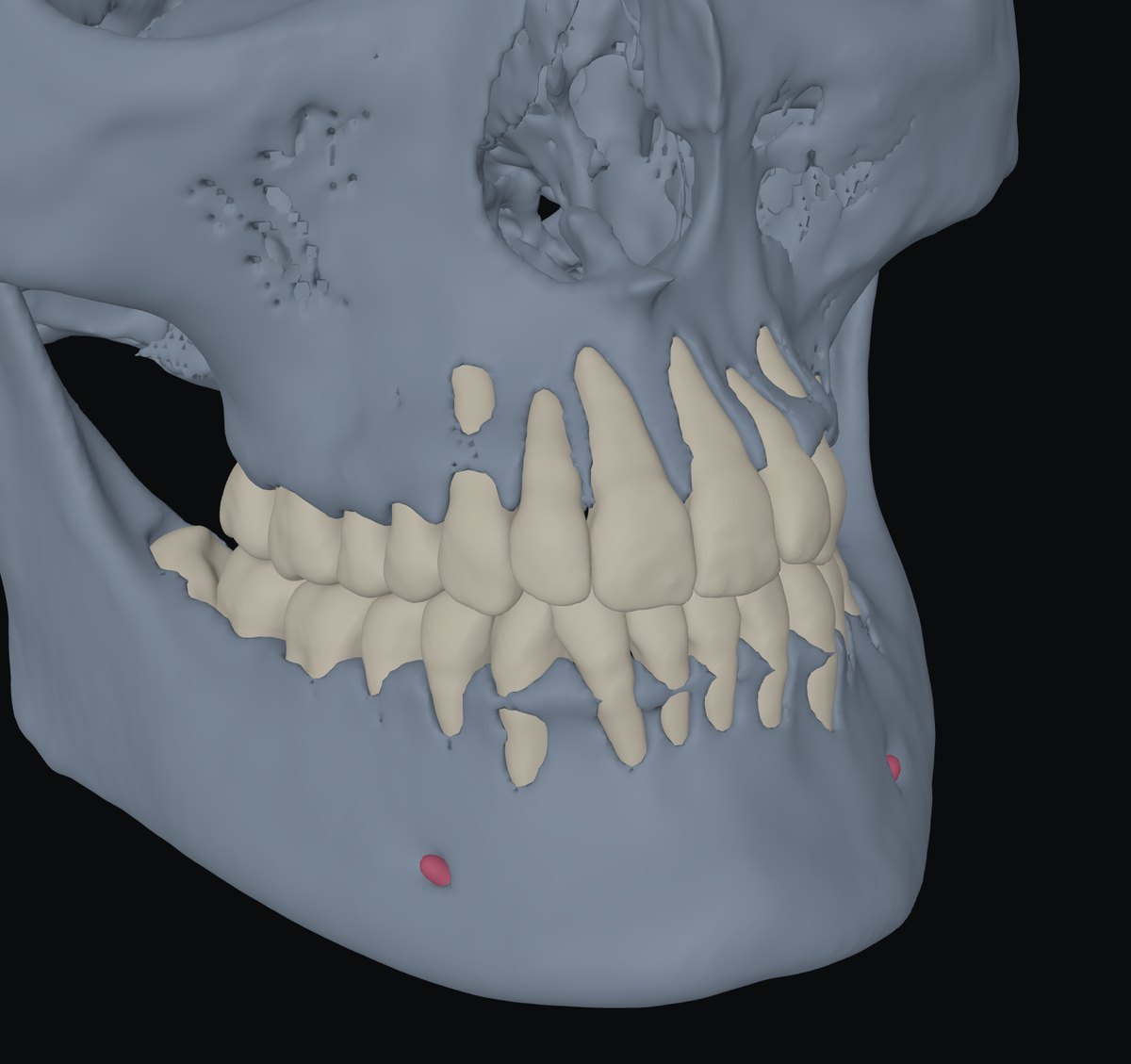
One button, Place scans: about a minute and a half per jaw.
- 2Scans land on the CT
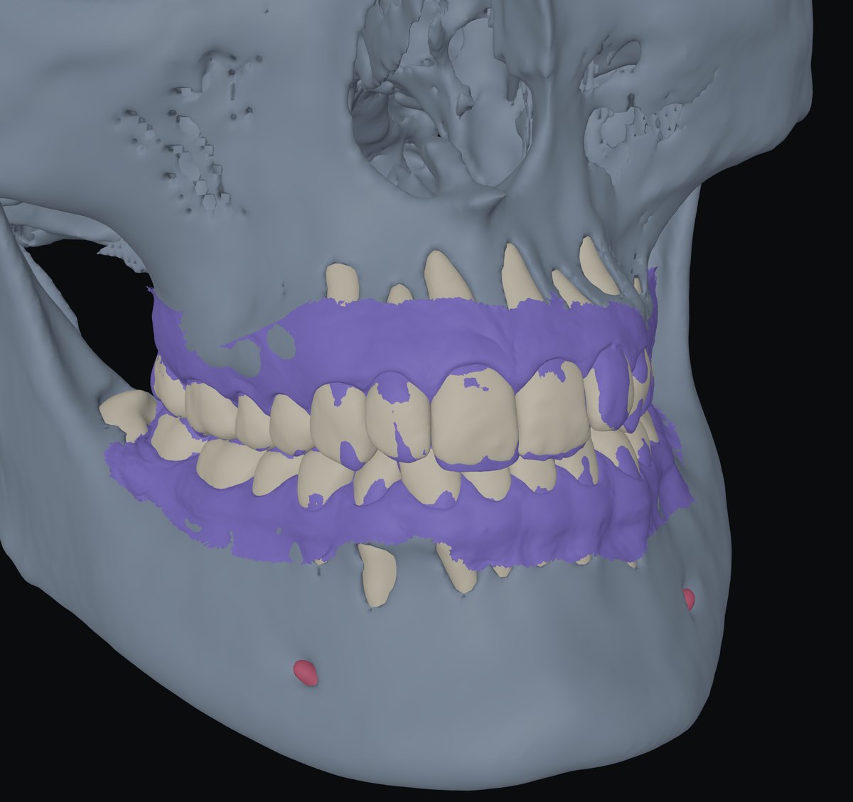
No manual points; each jaw is checked before it is shown.
- 3Crown + root per tooth
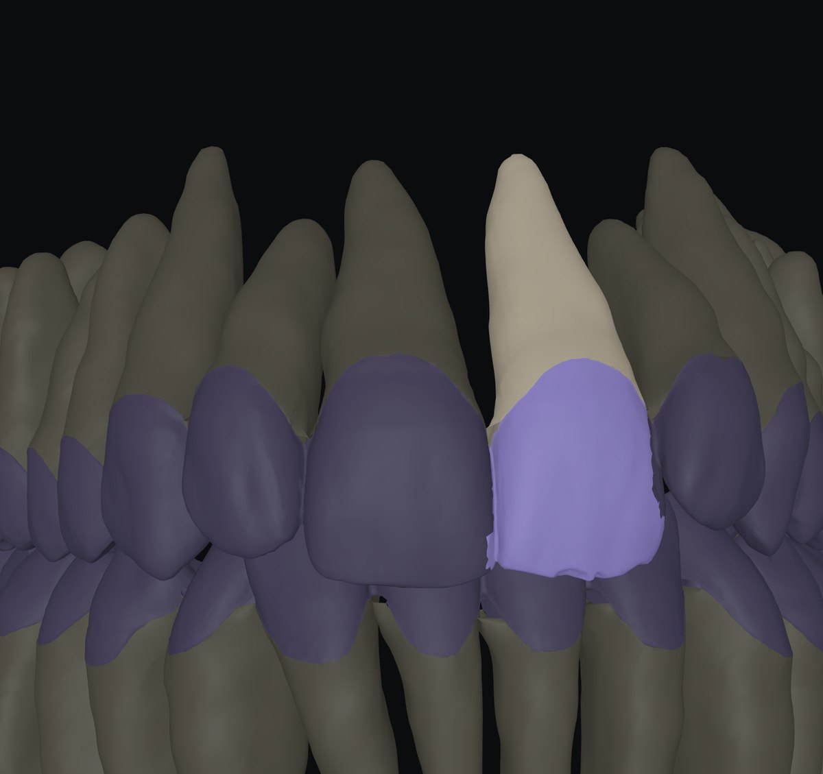
Crown from the scan, root from the CT; crowns and gum are exactly the whole scan.
What you getOn screen and to take away
- 3D of assembled teeth, the gum as its own layer, the scans on or off; crowns from the scan or from the CT.
- STL: tooth-NN-crown.stl, tooth-NN-root.stl, upper-gum.stl, upper-scan.stl… — all in the CT’s coordinates, with pose.txt.
- Share link with the placed scans.
Measured, not promisedOn our cases
- A pose is shown only after it passes a six-rule check; the check keeps the error under about 0.3°.
- Run again, the same pair lands within 0.02–0.12°. Placed by hand, within 0.2° of the automatic pose.
Not yet
- Check that the scans are this patient’s: the doctor confirms the result.
- Nothing is claimed below the gum line.
Next tool01 · Prep depth →
How much was removed — the pre-op scan over the prep, cut through, in millimetres.