04 / 05 · CT segmentationA DICOM folder, on a computer
CT segmentation
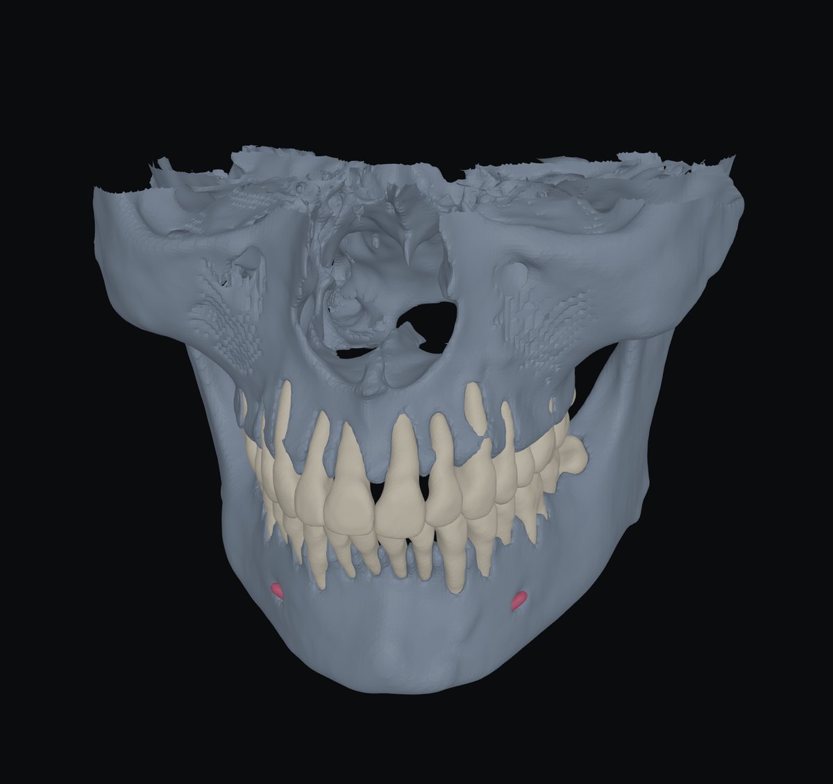
One CT into teeth by number, both jaws and the nerve canal — in 3D, as STL.
Where the canal runs under the roots of the 38 before an extraction, where an impacted canine sits against its neighbours — seen, not imagined from slices.
InDICOM folder
Out3D · STL · link
RunsBrowser + server in the EU
KeptNothing
How to use it3 steps · on a real case
- 1Choose the folder
Press Segment: the CT goes to a server in the EU without name or date; nothing is kept.
- 2Teeth, jaws, canal
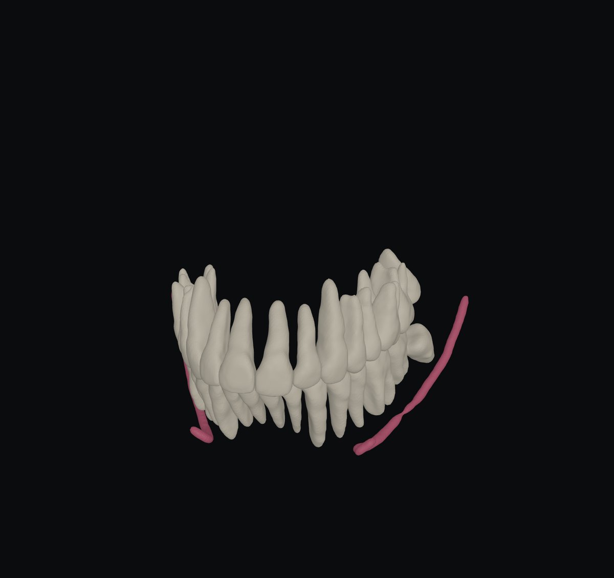
Each tooth by FDI number. Hide the bone, turn, point at a tooth.
- 3Fix, then export STL
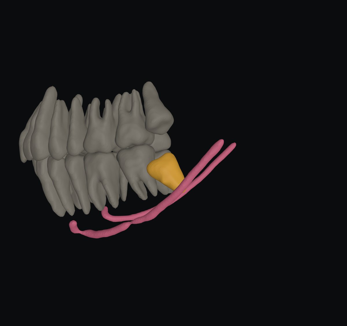
Reassign or split a suspicious piece; one STL per structure.
What you getOn screen and to take away
- 3D: every tooth with its FDI number, lower jaw, upper jaw, the mandibular canal; layers on and off.
- Fix: suspicious pieces listed — too big, on the wrong side of the bite, stray — click to make it bone, a tooth, or delete; a knife to split.
- STL: tooth-36.stl, mandible.stl, maxilla.stl, canal.stl — in the CT’s own coordinates.
- Share link: the 3D labels, without the CT.
Measured, not promisedOn our cases
Not yet
- A measuring tool, not a diagnosis.
Next tool05 · CT + scan →
The jaw scans placed on the CT: every tooth a crown from the scan and a root from the CT.